Hi everyone,
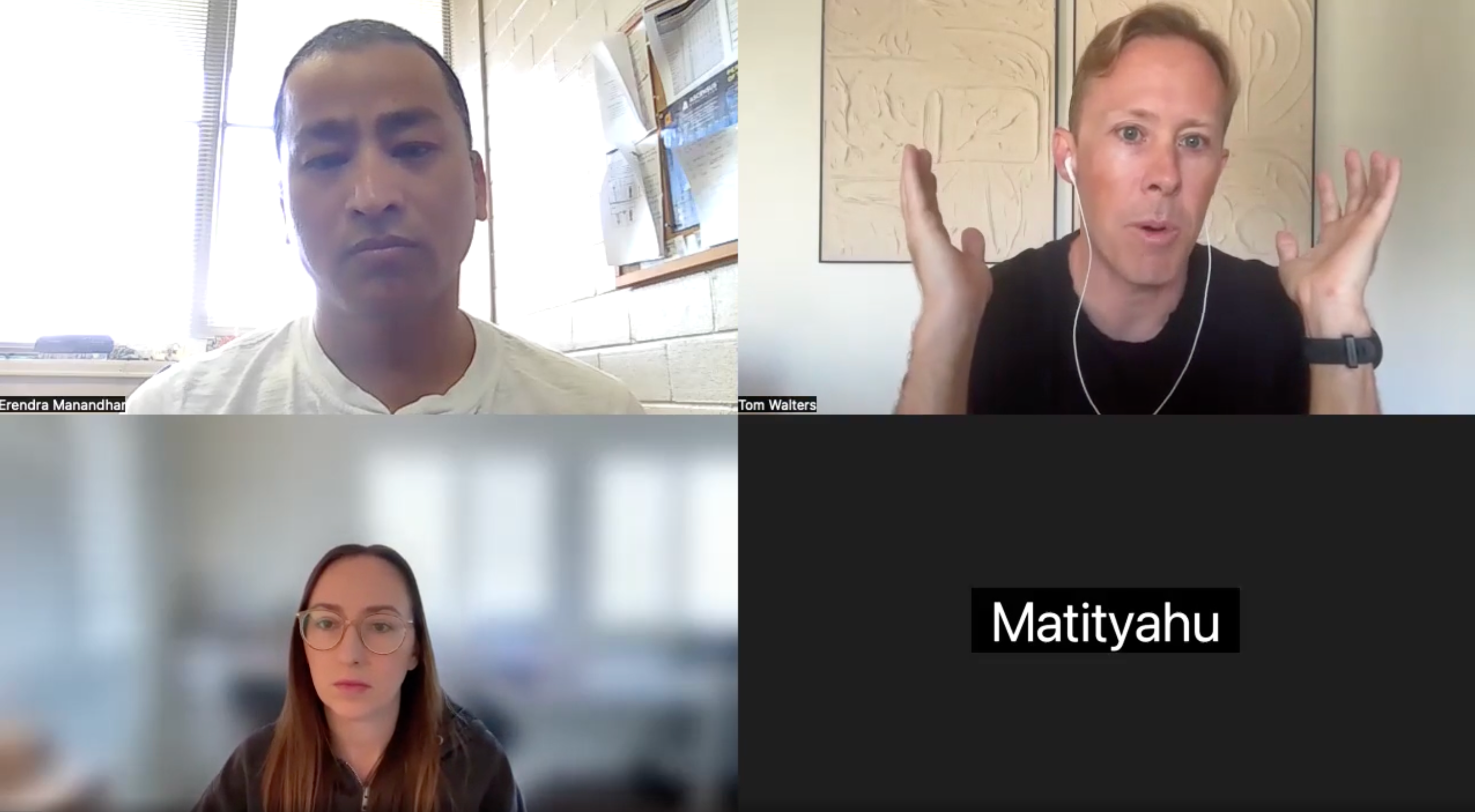
Here is a replay from today's office hours session. We discussed several issues, including cervical disc injuries, patellofemoral joint pain, IT band syndrome and nerve referral from the low back.

Hi everyone,
Here is a replay from today's office hours session. We discussed several issues, including cervical disc injuries, patellofemoral joint pain, IT band syndrome and nerve referral from the low back.
Hi everyone,
Our next office hours session will be this Friday at 10am PT. Please visit the link below to RSVP. This is your opportunity to ask me your specific rehab questions. I hope to see you there!
📚Osteoarthritis (OA) is degenerative condition of the cartilage present in our joints and most commonly affects the knee and hip.
🔎Symptoms usually include pain, swelling, joint stiffness, limited range of motion and difficulty with many functional tasks. Severe pain or functional loss associated with OA of the knee or hip often leads to joint replacement surgery.
🧠Many individuals with OA are told that exercise will harm their joint(s) and encouraged to avoid many activities. While it is important to respect one’s symptoms and progress gradually, it should be known that properly dosed exercise can actually improve the health of cartilage. Furthermore, strengthening the muscles that surround a given joint can actually help to reduce stress on cartilage and other sensitive structures.
✅A knee osteoarthritis program can be found here in the app. Here is a link for the program.
https://membership.rehabscience.com/programs/knee-osteoarthritis?category_id=258965
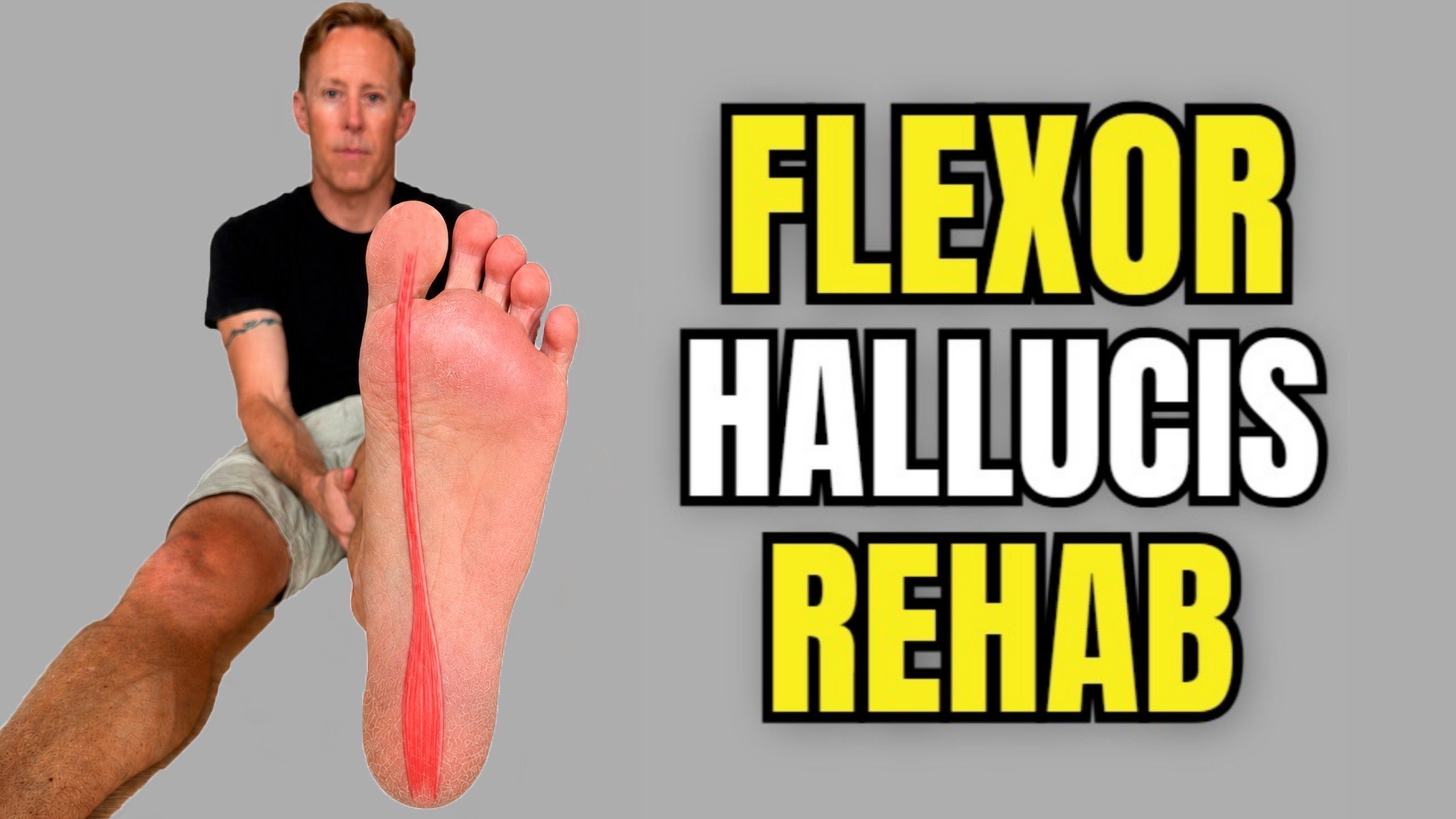
Hi everyone! A new video on Flexor Hallucis Longus (FHL) tendinopathy rehab is now available here in the app. If you have pain underneath your big toe, in the arch of your foot or on the inside of your ankle, these exercises are good to implement. Here is a link for the video.
👉Shoulder instability describes a condition of increased joint mobility that affects the static stabilizers of the shoulder joint (labrum, capsule and ligaments).
🔎When looking to improve shoulder stability, it’s important to perform exercises that target the four rotator cuff muscles and the other muscles that move and control the scapula (trapezius, serratus anterior, etc).
🧠Besides adding these types of exercises, it’s also important to consciously focus on activating the muscles that surround the shoulder joint when lifting the arm overhead or away from your body.
✅A more comprehensive program for shoulder instability can be found here in the app. Here is a link for the program.
https://membership.rehabscience.com/programs/shoulder-instability?category_id=259882
📚The neck or cervical spine includes seven vertebrae that are separated by six fibrocartilaginous intervertebral discs.
🧠The discs act as shock absorbers and also function as passive stabilizers of the spinal motion segment (2 vertebrae and the disc between them).
🔎This cervical fluoroscopy showcases flexion and extension of the cervical spine and is a great visual tool for observing our anatomy in action!
📚The anterior cruciate ligament (ACL) runs obliquely from the anterior intercondylar area of the tibia up to the posteromedial aspect of the lateral femoral condyle, and plays a critical role in preventing anterior translation of the tibia relative to the femur.
🔎In this dissection, the femoral condyles roll posteriorly over the tibial plateau as the knee joint flexes, allowing the intercondylar notch to open and reveal the ACL.
🧠Visualizing the ACL during flexion highlights its functional orientation and importance in dynamic knee stability. As the femur moves on the tibia, the ACL becomes taut, particularly in mid-to-deep flexion, helping to resist excessive anterior glide and rotational forces.
✅If you need a rehab program for an injury of the ACL or another ligament in the knee, the knee ligament injury program here in the app will teach you some of the most important exercises. Here is a link for this program.
https://membership.rehabscience.com/programs/knee-ligament-injury?category_id=258965
📚The IT band (ITB) is a dense, fibrous band of connective tissue that runs along the lateral thigh from the hip to the knee. ITB syndrome typically manifests as lateral knee pain (see image) and is one of the most common overuse injuries occurring in 12-25% of runners and 12.5% of cyclists.
🧠While stretching may help temporarily reduce pain in some people, research suggests that affected individuals should focus on incorporating exercises that strengthen the hip ABDuctors and motor control exercises designed to prevent the affected limb from deviating away from neutral when standing on one leg as this may sensitize the region.
🔎In general, 3 sets of 8-12 repetitions of each exercise should be performed 2-3 times per week. On the standing exercises, focus on keeping the knee aligned over the foot.
✅The app includes a more comprehensive IT band program that guides you through three phases of rehab. Click the link below to access the IT band syndrome program.
https://membership.rehabscience.com/programs/iliotibial-band-pain?category_id=258965
📚The fibularis muscles (fibularis longus & fibularis brevis) run along the lateral lower leg and serve as the primary evertors of the ankle.
🔎Besides everting the ankle and assisting in plantarflexion, the fibularis muscles also help stabilize the ankle during standing tasks, especially when on one leg or navigating uneven surfaces.
🧠During more severe inversion ankle sprains, the fibularis muscles (along with the lateral ankle ligaments) can be stretched and injured.
👉If you have pain in this region or have suffered a recent ankle sprain, the massage technique shown here may help reduce pain and improve function.
✅Besides trying this technique, you would also want to add exercises that strengthen these muscles. The ankle sprain program here in the app will teach you exercises for these muscles. Here is a link for the ankle sprain program.
https://membership.rehabscience.com/programs/ankle-sprain?category_id=261017
🔎Here is a great dissection that shows how the gastrocnemius and soleus muscles blend into the Achilles tendon and how the tendon is influenced by ankle movement.
🧠During ankle dorsiflexion, we see how the Achilles tendon and calf muscles are put on tension. Then, when the ankle moves into plantarflexion, the tendon and muscles are put on slack.
✅If you are suffering from Achilles tendon issues or a calf strain, I would recommend following the calf and Achilles pain program here in the app. Here is a link for the program.
https://membership.rehabscience.com/programs/achilles-tendinopathy?category_id=261017