Tom W.

Image for the badge Activation.
Activation earned for a 1-day streak!
Image for the badge Control.
Control earned for a 7-day streak!

Posted

30 May 13:52

00:16

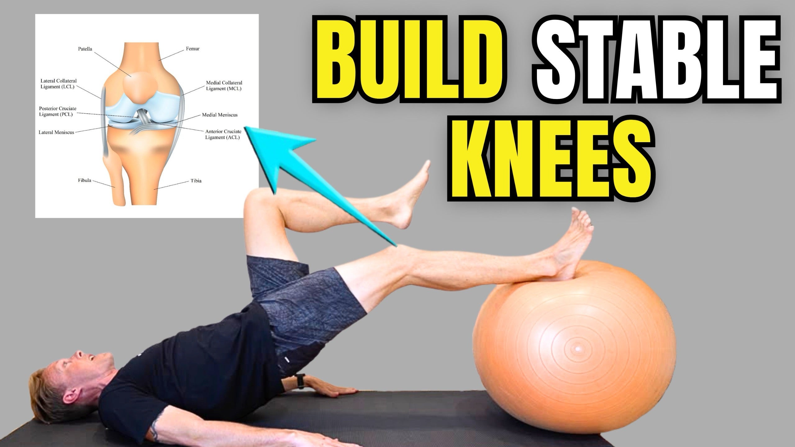
📚The inside of the knee is stabilized by a group of structures called the medial knee complex, with two key ligaments being the MCL and POL.

MCL (Medial Collateral Ligament)

🔎The MCL is a broad ligament that runs from the femur to the tibia along the inner knee. Its main job is to resist forces that push the knee inward (valgus stress).

It has:

• Superficial MCL → primary restraint to valgus stress

• Deep MCL → blends with the medial meniscus and joint capsule for added stability

POL (Posterior Oblique Ligament)

🔎The POL sits just behind the MCL in the posteromedial corner of the knee. It works with the joint capsule and semimembranosus tendon to stabilize the knee near full extension.

Its main roles include controlling:

• Tibial rotation

• Valgus stress in extension

• Posteromedial knee stability

✅Together, the MCL and POL help control side-to-side movement and rotation of the knee during walking, cutting, pivoting, and sport activities.

Posted

28 May 10:56

Posted

26 May 12:34

00:56

Hi everyone, I know many of you already have my big red book, but I wanted to let you know that my new smaller body region specific books were released today!

If you’re looking for a smaller book that is easier to carry when traveling or going to the gym, these books would be a great option.

The Amazon link below will allow you to view all of the new books.

https://www.amazon.com/stores/Tom-Walters/author/B0BC4N3RWG?ref=ap_rdr&shoppingPortalEnabled=true

Posted

26 May 12:22

00:29

👉Posterior to anterior or PA spinal mobilizations are one of my favorite techniques for treating patients with unilateral or one-sided back pain.

🔎When performing this technique, I start at the bottom of the lumbar spine (L5-S1) and work up one segment at a time until I reach the bottom of the thoracic spine (T12).

🧠This is done first as an assessment to determine if the patient’s symptoms can be reproduced with the mobilization pressure/movement. In many cases, familiar pain is reproduced at one or two spinal levels and then those levels are mobilized further.

✅When mobilizing (checkout the Thumb Saver tool to protect your thumbs) a painful level, small oscillations are delivered to the painful area at an intensity that is perceived to be therapeutic by the patient. I will usually perform the mobilization for 1-2 minutes and perform 3-5 sets of the technique.

Posted

23 May 09:46

01:00

🔎The iliacus and psoas major muscles are our primary hip flexors. Psoas major is the deepest core muscle and the only muscle that attaches the trunk to the lower limb. It runs from the vertebrae and discs of the low back (T12-L5) down to the lesser trochanter on the inside of the femur. This unique configuration allows psoas to possess the dual role of spinal stabilizer and hip flexor.

🧠If you are experiencing pain in the low back, sacroiliac joint region or hip, it could help to address your hip flexors. The exercises in this video are some of the most helpful.

✅A more comprehensive hip flexor rehab program can be found here in the app under the hip category. Here is a link for the program.

https://membership.rehabscience.com/programs/hip-flexor-pain?category_id=258455

1️⃣Hip Flexor Stretch: Place the leg you would like to stretch down. Before moving from this position, make sure to squeeze your abs and glutes to place the pelvis in a position of posterior tilt, which help take any slack out of the hip flexors. Lunge forward slightly without arching the low back. Hold for 15-30 seconds and repeat 2-4 times each day.

2️⃣Front Plank: The front plank creates an isometric hip flexor contraction and is a great place to begin training if have had a recent injury. Shoot for 3 repetitions and hold each one for 20 seconds.

3️⃣Hip Flexor March: Place a mini-band around your feet and flex the affected hip as far as possible. Feel free to hold onto something, if you need to, as this is not necessarily meant to be a balance training exercise. Perform 3 sets of 12-15 reps.

Posted

22 May 09:30

Image from Tom W.'s post

Here is a link for our next office hours session. Please click the link to RSVP, so that I know you are planning to attend. 

https://membership.rehabscience.com/programs/live-2yflubkzbge?utm_source=product&utm_medium=share&utm_campaign=content_share&utm_term=4204755&utm_content=copy

5

Posted

21 May 11:32

Image from Tom W.'s post

Hey everyone! I published a new video this morning on glute exercises that can help reduce pain and improve gait mechanics. Here is a link for the video.

https://membership.rehabscience.com/programs/glute-exercises-for-gait?utm_source=product&utm_medium=share&utm_campaign=content_share&utm_term=4202791&utm_content=mobile

Posted

21 May 08:04

00:04

👉Did you know that most (66.66%) disc herniations reabsorb with time and conservative care.

🧬The images in this post are from a case report from the New England Journal of Medicine (see reference) that documents the spontaneous resorption of a disc herniation without surgery.

🔎Case Study: A 29-year-old woman presented to the spine clinic with new-onset pain in her right leg, accompanied by paresthesia. MRI of the lumbar spine revealed a lumbar disc herniation resulting in substantial spinal stenosis and nerve-root compression (first image).

📗She elected conservative treatment with physical therapy and an epidural injection of glucocorticoids. A second MRI obtained at follow-up 5 months after presentation showed resolution of the herniation (second image). Her clinical symptoms resolved, and she was discharged from the clinic.

🧠The details of this individual’s rehab were not given in the case study, but it is important to know that relatively simple programs that include movements like walking, general low back mobility drills and low-load resistance training exercises help the majority of people.

✅A program for disc herniations can be found in the back and spine section here in the app.

📘References:

1. Hong J and Ball PA. Resolution of Lumbar Disk Herniation without Surgery. N Engl J Med. 2016.

2. Zhong M, et al. Incidence of Spontaneous Resorption of Lumbar Disc Herniation: A Meta-Analysis. Pain Physician. 2017.

3. Chiu C, et al. The probability of spontaneous regression of lumbar herniated disc: a systematic review. Clin Rehabil. 2015.

Posted

14 May 11:55

00:22

👉The rhomboid and trapezius muscles are located between the spine and shoulder blades and play a role in neck, back and shoulder movement health.

🔎Today’s post shows how the rhomboids (left side) and trapezius (right side) can work to pull the scapula (shoulder blade) backwards (retraction) toward the spine.

🧠Pain in this intrascapular region is quite common and can be associated with the muscles in the region, the underlying thoracic and rib joints, or can be referred from the neck.

✅If you are experiencing pain in this region, I would recommend implementing the exercises in the thoracic pain program here in the app. Here is a link for the program.

https://membership.rehabscience.com/programs/thoracic-pain?utm_source=product&utm_medium=share&utm_campaign=content_share&utm_term=3788553&utm_content=mobile

3

Posted

13 May 10:14