Sorted by
Newest posts Popular Latest activity
 
May 13
Image from Tom W.'s post
 
May 10
00:16

📚The anterior compartment of the forearm contains the wrist and finger flexors, which are responsible for flexing the wrist and providing grip strength.

🧠Soft tissue mobilization (massage) of these muscles is often implemented in physical therapy and can help with conditions like golfer’s elbow (medial epicondylalgia), carpal tunnel syndrome and various wrist pain issues.

🔎To mobilize these muscles, work from the distal forearm toward the inside of the elbow (medial epicondyle). Starting all the way down at the wrist usually isn’t necessary or comfortable as the majority of the muscle is located more proximally.

✅The app includes a comprehensive golfer’s elbow program that shows how to perform these types of soft tissue mobilizations on yourself. Visit the elbow section to find this program.

2
 
May 09
00:43

📚The suboccipital muscles are a small but incredibly important group of muscles located at the base of the skull. These four muscles connect the upper cervical spine (C1 and C2) to the occiput and help control the fine movements of the head and neck.

🔎Their primary roles include subtle head rotation, extension, and stabilization, but they also contain a very high density of sensory receptors. This means they play a major role in proprioception, balance, eye movement coordination, and helping your brain understand where your head is positioned in space.

👉The four suboccipital muscles include:

• Rectus capitis posterior major  

• Rectus capitis posterior minor  

• Obliquus capitis superior  

• Obliquus capitis inferior

🧠Because these muscles work constantly to stabilize the head, they can become overactive or sensitive with prolonged screen time, stress, forward head posture, headaches, or neck injuries. Tightness and irritation in this region are commonly associated with cervicogenic headaches and upper neck pain.

✅Improving upper cervical mobility, posture awareness, breathing mechanics, and deep neck muscle function can often help reduce excessive strain in this area.

2
 
May 08
00:09

📚Shoulder abduction isn’t just the arm lifting out to the side, it’s a coordinated effort across multiple joints.

🔎The movement starts at the glenohumeral joint, where the supraspinatus initiates abduction and the deltoid drives it. At the same time, the rotator cuff stabilizes the humeral head to prevent excessive upward movement.

🧠As the arm continues upward, the scapula joins in. This creates the classic 2:1 rhythm, meaning for every 3° of motion, 2° comes from the shoulder joint and 1° from the scapula.

👉This scapular motion is controlled by the trapezius and serratus anterior, helping rotate the scapula upward and clear space for the arm to move overhead.

✅Smooth shoulder motion is less about one muscle and more about how everything works together.

 
May 06
Image from Tom W.'s post
 
May 04
Image from Tom W.'s post

Hi everyone, our next office hours session will be this Wednesday at 9am PT. Here is a link to RSVP. I hope to see you there!

https://membership.rehabscience.com/programs/live-tdjv-ogkgoe?category_id=262577&utm_source=product&utm_medium=share&utm_campaign=content_share&utm_term=4178799&utm_content=copy

 
May 02
00:27

🧠Pain in the muscles along the medial tibial border can occur with posterior shin splints, calf strains, tibialis posterior tendinopathy and arch related issues, like plantar fasciopathy.

🔎If you are having pain in this region, the massage technique shown in this video may be helpful. It is an area I often find myself working when patients present with the conditions mentioned above.

✅Besides soft tissue/massage work, make sure to also add the exercises for the calf muscles and tibialis posterior. The shin splints program here in the membership is a good place to start.

 
April 29
Image from Tom W.'s post
 
April 27
• Edited (Apr 27, 2026)
00:08

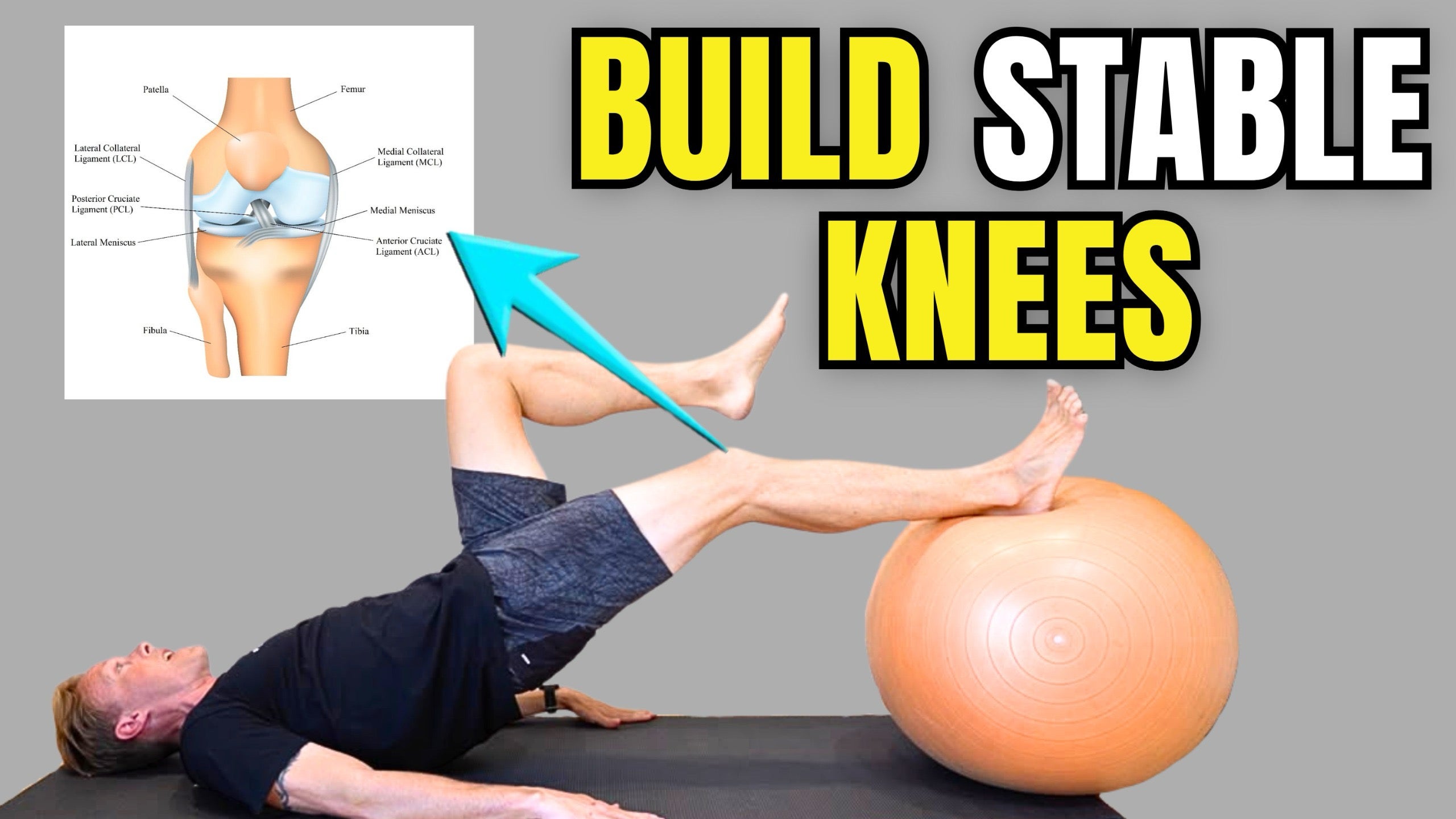
📚The ACL connects the femur (thigh bone) to the tibia (shin bone) and runs at an oblique angle from the posterior aspect of the femur to the anterior aspect of the tibia. Due to this arrangement, the ACL is responsible for preventing anterior translation of the tibia or posterior translation of the femur.

🧠In the anterior drawer test, which is being demonstrated in the first video, the examiner attempts to pull the tibia anteriorly. If the ACL is compromised, the tibia will translate forward farther than it normally should.

✅Here in the app you will find a comprehensive rehabilitation program for knee ligament injuries, which can be found under the knee section. Here is a link for that program.

https://membership.rehabscience.com/programs/knee-ligament-injury?utm_source=product&utm_medium=share&utm_campaign=content_share&utm_term=3776140&utm_content=mobile

 
April 23
Image from Tom W.'s post